
INTRODUCTION
Mid-portion (or non-insertional) Achilles tendinopathy has been reported as one of the most common overuse injuries (Maffulli et al., 2003). It is common in those who engage in regular physical activity, which means athletes are particularly susceptible to this condition. Sports physiotherapists who treat regularly treat runners will be aware of its high incidence within this population. It is, however, common in many sports (Carcia et al., 2010). This highlights the importance that sports physiotherapists are aware of the most evidence based methods of assessment, diagnosis and management.
This post, you may notice, reflects the evidence discussed in clinical practice guidelines and systematic reviews on the Achilles tendinopathy (Carcia et al., 2010; Magnussen et al., 2009). Enjoy!
RISK FACTORS FOR ACHILLES TENDINOPATHY
The literature identifies a number of risk factors for the development of Achilles tendinopathy. These include:
- Abnormal dorsiflexion range of motion (Kaufman et al., 1999)
- Abnormal subtalar joint range of motion (Kaufman et al., 1999; Kvist et al., 1991)
- Plantarflexor weakness (McCrory et al., 1999)
- Overpronation (McCrory et al., 1999)
- Changes in training habits; increases, surface changes, return from break
- Poor equipment e.g. footwear
It is essential that as sports physiotherapists we are aware of these risk factors of developing Achilles tendinopathy, as frequently they will guide decisions regarding both prevention and treatment.
DIAGNOSIS OF ACHILLES TENDINOPATHY
SUBJECTIVE EXAMINATION: The athlete will often report:
- Pain localised to the Achilles tendon region
- Intermittent pain associated with exercises/activity (Maffulli & Kader, 2002)
- “Start-Up” Pain i.e. painful at beginning of exercise, which settles when warmed up (Leach et al., 1981)
- Achilles tendon stiffness, particularly following rest.
OBJECTIVE EXAMINATION:
- Observation: often thickening or swelling of the tendon visible. Foot posture is also an important consideration.
- AROM: Frequently decrease in dorsiflexion range of motion (Kaufman et al., 1999)
- MMT: Weakness and reproduction of pain with plantarflexion
- Palpation: Achilles Tendon Palpation Test (i.e. tenderness 2-6cm from insertion) has been shown to have a sensitivity of 58% and specificity of 84% (Maffulli et al., 2003). Tautness and active trigger points in the calf muscle bellies may also be palpated.
- Arc Sign: Sensitivty of 52% and specificity of 83%.
NB: As well as making a clinical diagnosis of Achilles tendinopathy, it is evident that during the physical or objective examination that you should identify any abnormalities or contributors (biomechanical or otherwise) to the condition amenable to physiotherapy interventions e.g. overpronation.
DIFFERENTIAL DIAGNOSES
This is a list of potential differential diagnoses for the posterior ankle pain (Carcia et al., 2010):
- Partial or Complete achilles tendon tear
- Posterior Ankle Impingement
- Sural Nerve Pathology
- Os Trigonum Syndrome
IMAGING
Imaging is required when your examination findings do not allow for a clear diagnosis, or to exclude competing hypotheses such as those above. Ultrasound and MRI are the two options for imaging the achilles tendon, the diagnostic accuracy of which is shown below (Khan et al., 2003):

Diagnostic Imaging For Achilles Tendinopathy
TREATMENT FOR ACHILLES TENDINOPATHY
PHYSIOTHERAPY MANAGEMENT OF ACHILLES TENDINOPATHY
There are a number of conservative measures, I’m sure you are aware, for the management of Achilles tendinopathy. It has been suggested that a 4 – 6 month trial of conservative management be undertaken prior to any surgical management (Johnston et al., 1997). The physiotherapy management of Achilles tendinopathy may include:
Eccentric Loading (Carcia et al., 2010; Magnussen et al., 2009): this seems to be the cornerstone of physiotherapy management on non-insertional Achilles tendinopathy. It has been evaluated and proven effective in many studies. However, the most appropriate prescription and population likely to respond to eccentric loading is yet to be investigated.
Stretching – of both the gastrocnemius and soleus (Park et al., 2006)
Manual Therapies, likely including:
- Soft tissue massage/myofascial techniques
- Talocrural mobilisation: commonly T/C AP’s to normalise dorsiflexion range of motion
- Subtalar Mobilisation: To normalise sub-talar joint range of motion
Taping: most comonly this would include low dye taping to assist with prevention of overpronation during gait and running (Carcia et al., 2010). However, you may also consider the less evidence based taping techniques including kinesiotaping or dynamic taping.
Foot Orthoses: orthotics have been shown to reduce pain and improve foot kinematics during running in patients with achilles tendinopathy (Mayer et al., 2007). They should be considered in the management of patients with achilles tendinopathy.
Low Level Laser Therapy: Stergioulas et al. (2008) evaluated the addition of low level laser therapy to an eccentric exercise program and found favourable outcomes in terms of pain and stiffness, and therefore should be considered in patients with achilles tendinopathy.
NON-PHYSIOTHERAPY CONSERVATIVE MANAGEMENT OF ACHILLES TENDINOPATHY
Given the failure of physiotherapy management you may suggest the trial of a few other conservative techniques to assist in the management of Achilles tendinopathy. These include:
Platelet-Rich Plasma Injections: there has been some conflicting evidence on the use of PRP (click the link). Gaweda et al. (2010) found a significant improvement in symptoms of Achilles tendinopathy using PRP injections.
Topical Glycerol TriNitrate (GTN): Paoloni et al. (2004) evaluated the use of topical GTN patches, combined with an eccentric exercise program, and found they decreased pain in the treatment group. GTN patches can therefore be utilised to complement an exercise program.
Steroid Injections: A recent systematic review by Magnussen et al. (2009) found conflicting evidence on the use of steroidal injections in the management of achilles tendinopathy, and could not recommend their use on the current evidence base.
SURGICAL MANAGEMENT OF ACHILLES TENDINOPATHY
Surgical management is only indicated in the recalcitrant cases of Achilles tendinopathy i.e. following the failure of physiotherapy and other invasive management techniques. The common surgical techniques utilised and described are (Murphy, 2009):
- Tenotomy
- Debridement
- Repair
It is challenging to make evidence based comparisons between the surgical techniques. The overall long-term results of surgical management of Achilles tendinopathy are positive, with articles citing “satisfactory” results in about 79 – 85% of cases (Leach et al., 1992; Schepsis et al., 1994). However, it should be noted that there is frequently a high recurrence or deterioration rate in some athletes, particularly runners, and some require further surgery (16%) (Schepsis et al., 1994).
TAKE HOME MESSAGES
- Diagnosis of Achilles tendinopathy should be based on a combination of subjective, objective and imaging (as required) findings
- Eccentric exercises are go
- Effective physiotherapy management requires identification of kinetic chain/biomechanical contributors
- Non-physiotherapy/Invasive techniques can complement your physiotherapy management
- Surgery is only indicated in the very recalcitrant cases (i.e. recommended 4 – 6 months of conservative management)
What are your beefs with Achilles tendinopathy, and the evidence base for treatment (I’m sure there are plenty out there)? Be sure to let me know in the comments or catch me on Facebook or Twitter
If you require any sports physiotherapy products be sure check out PhysioSupplies (AUS) or MedEx Supply (Worldwide)
REFERENCES
Gaweda, K., Tarczynska, M., Krzyzanowski, W.Treatment of Achilles Tendinopathy with Platelet-Rich Plasma. Int J Sports Med 2010; 31: 577-583
Johnston E, Scranton P, Jr., Pfeffer GB. Chronic disorders of the Achilles tendon: results of conservative and surgical treatments. Foot Ankle Int. 1997;18:570-574
Kaufman KR, Brodine SK, Shaffer RA, Johnson CW, Cullison TR. The effect of foot structure and range of motion on musculoskeletal overuse injuries. Am J Sports Med. 1999;27:585-593
Khan KM, Forster BB, Robinson J, et al. Are ultrasound and magnetic resonance imaging of value in assessment of Achilles tendon disorders? A two year prospective study. Br J Sports Med. 2003;37:149-153
Kvist M. Achilles tendon injuries in athletes. Ann Chir Gynaecol. 1991;80:188-201
Leach, Robert E.; Schepss, Anthony A.; Takai, Hiroaki Long-Term Results of Surgical Management of Achilles Tendinitis in Runners. Clinical Orthopaedics & Related Research 1992;282:208-212
Leach RE, James S, Wasilewski S. Achilles tendinitis. Am J Sports Med. 1981;9:93-98
Mafulli N, Kader D. Tendinopathy of tendo achillis. J Bone Joint Surg Br. 2002;84:1-8.
Mafulli N, Kenward MG, Testa V, Capasso G, Regine R, King JB. Clinical diagnosis of Achilles tendinopathy with tendinosis. Clin J Sport Med. 2003;13:11-15.
Mafulli N, Wong J, Almekinders LC. Types and epidemiology of tendinopathy. Clin J Sports Med. 2003;22:675-692
Magnussen RA, Dunn WR, Thomson AB. Nonoperative Treatment of Midportion Achilles Tendinopathy: A Systematic Review. Clinical Journal of Sport Medicine. 2009;19(1): 54-64
Mayer F, Hirschmuller A, Muller S, Schuberth M, Bauer H. Effects of short-term treatment strategies over 4 weeks in Achilles tendinopathy. Br J Sports Med. 2007;41:e6
McCrory JL, Martin DF, Lowery RB, et al. Etiologic factors associated with Achilles tendinitis in runners. Med Sci Sports Exerc. 1999;31:1374-1381.
Murphy GA. Surgical treatment of non-insertional Achilles tendinitis. Foot and Ankle Clinics 2009; 14(4); 651-661.
Paoloni J, Appleyard R, Nelson J, et al. Topical glyceryl trinitrate treatment ofchronic noninsertional Achilles tendinopathy. J Bone Joint Surg 2004;86-A:916–21
Schepsis AA, Wagner C, Leach RE. Surgical Management of Achilles Tendon Overuse Injuries: A Long-term Follow-up Study Am J Sports Med 1994;22: 611-619
Stergioulas A, Stergioula M, Arrskog R, Lopes-Martins RA, Bjordal JM. Effects of low-level laser therapy and eccentric exercises in the treatment of recreational athletes with chronic achilles tendinopathy. Am J Sports Med. 2008;36;881-887.
Related Posts
 Lateral Epicondylalgia (Tennis Elbow): Evidence Based Assessment and Management
 Athletic Pubalgia (Sports Hernia): Evidence Based Diagnosis and Management
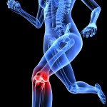 Patellar Tendinopathy: The Efficacy of Injection Treatments
 Kienbock’s Disease: Evidence Based Assessment and Management
 Does A Forefoot Strike Pattern Reduce Injury?
 The Running Shoe: Excellent Science or Excellent Marketing?
Comments
Trackbacks
-
Achilles Tendinopathy Evidence Based Diagnosis And Management…
[…]The literature identifies a number of risk factors for the development of Achiles tendinopathy. These include: Abnormal dorsiflexion range[…]…
You can think about radial shockwave therapy for achilles tendinitis.It does help the patient in terms of pain and movement.Once better with the pain ,then we can start with some gentle stretches and then progress to eecentric exercises.
Thanks for your input Anil, and good point, you can hear more about Achilles tendinopathy from Prof. Karim Khan in his interview about Achilles Tendinopathy.
I have had great success in eliminating “achilles tendonitis”, for many years by not treating the achilles area at all. Instead I treat L5S1 by continuously mobilising unilateraly there, untill tone has reduced and passive movements there are no longer able to elicit pain local the the facet joint. By doing so I restore a non threatened state of mobility to that portion of the lumbar spine most closely associated with the sciatic nerve.
Achilles symptoms reduce as spinal protective behaviour reduces . One to three treatments are usually needed to fully eliminate all signs and symptoms at the achilles.
Indeed I don’t recall a single incident where this treatment has not brought about rapid and lasting resolution at the achilles in more than twenty years of private practice.
I think what does often happen when therapists do pay appropriate attention with treatment at the lower lumbar spine in these cases, is that the period of mobilisation is too short. I commonly will approach and continuously mobilise (in cases of achilles symptoms) both sides and include L4, most importantly, till protective tone has normalised. This may take up to ten minutes in some cases.
Cheers