
Introduction
In the world of sports physiotherapy and sports medicine articular cartilage defects of the knee are commonly seen (Reinold et al., 2006). Unfortunately, in these cases we find non-operative approaches are ineffective given the avascular nature of articular cartilage. Thus, there has been the development of a large number of surgical techniques to address articular cartilage lesions. This article will discuss a relatively new technique, Matrix-induced autologous cartilage implantation or MACI, including an overview of technique, mid-term outcomes and of course the rehabilitation implications for physiotherapists.
What Is Matrix-Induced Autologous Cartilage Implantation (MACI)?
As you are aware, damaged hyaline cartilage does not heal. When subchondral bone is also damaged some healing may take place via the formation of fibrocartilage (i.e. the basis for surgical “repair” techniques of microfractures and abrasion chondroplasty). However, given the inferior biomechanical properties of fibrocartilage in comparison to hyaline cartilage this has lead to variable clinical outcomes, particularly in the instance of large chondral defects (Peterson et al., 2002).
This has lead to the advent of a new surgical technique for large chondral defects of the knee which can create hyaline-like cartilage with good mechanical properties and contains normal cartilage components (Enea et al., 2011). The technique of matrix-induced autologous chondrocyte implantation (MACI) involves two surgical procedures. The first involves arthroscopic debridement of the lesion back to a stable edge and an initial harvest of healthy cartilage, the chondrocytes of which are then isolated and expanded ex-vivo. These new chondrocytes are then seeded directly onto a collagen membrane. The second surgery (generally months later and via arthrotomy) involves the re-implantation into the knee as a cell-scaffold construct and held in place with a fibrin glue. Then the surgeon hands them over to us, and thus begins the long and arduous rehabilitation process.
MACI – A Video Explanation
Physiotherapy Rehabilitation and Post-operative Management
Before I discuss anything, if you are regularly undertaking rehabilitation of these surgeries or have a particular interest in MACI surgery then you should take the time to access and read the articles listed below. They have true implications for physiotherapists, and are the core pieces of literature on which the rehabilitation programs below are based.
- Reinold MM, Wilk KE, Macrina LC, Dugas JR, Cain EL. Current Concepts in the Rehabilitation Following Articular Cartilage Repair Procedures in the Knee. J Orthop Sports Phys Ther 2006;36(10):774-794.
- Hambly K, Bobic V, Wondrasch B, Van Assche and Marolvits S. Autologous Chondrocyte Implantation Postoperative Care and Rehabilitation. American Journal of Sports Medicine 2006;34:(6);1020-1038
The first thing to note when undertaking a post-operative rehabilitation following a MACI procedure is that all patients are individuals! Now this may seem simple but it is essential to note that no two injuries, patients or surgeries are the same. It is important to take into account 4 key factors, and individualise your programs accordingly (Reinold et al., 2006). These are:
- Lesion: which includes location, size, depth, containment, quality of surrounding tissue
- Patient: which includes age, BMI, general health, activity level, goals, motivation, quality of cartilage
- Surgery: which includes repair procedure, tissue involvement, any concomitant procedures
- Surgeon: each surgeon will have their own preferences when it comes to rehabilitation
It is also worth noting what we are trying to achieve during the process of rehabilitation. Our goals of management throughout the rehabilitation process are:
- Protect the repair site by avoiding potentially deleterious forces and controlling load application
- Create a healing environment: with appropriate loads and knee mobilisation
- Reduce pain and effusion
- Restore soft-tissue balance: maintain mobility in relevant tissues
- Restore muscular function: consider the entire kinetic chain
- Enhance proprioception and neuromuscular control: particularly important in later stages
- Optimise overall athletic performance in late stage rehabilitation
As stated previously, there are large individual differences between different sites of repair. One major difference is that between a patellofemoral (i.e. patella or trochlea) and tibiofemoral repair. Thus, separate rough rehabilitation guidelines will be provided for these two repairs. Furthermore, it is worth noting that these programs are based on older literature and newer articles touting the advantages of accelerated programs have been published more recently (Ebert et al., 2008). Whilst the longer term results of these accelerated rehabilitation programs are not fully understood different surgeons seems to be using combinations of the 2 protocols. Anyways get the message: all patients, repairs, and orthopods will be different!
Post-Operative Physiotherapy Rehabilitation Following Patellofemoral MACI
In order to most effectively rehabilitate lesions of the patellofemoral joint you must have a superior knowledge of the biomechanics of the patellofemoral joint (which you no doubt will all have). This will allow you to individualise your program to the patient and repair site to load or unload the site in degrees of knee flexion. Quickly brush up on your biomechanics here. Now, to our rough rehabilitation guideline. It is worth noting that any progression through the phases requires tolerance of the previous activities/exercises and that any increases in pain, effusion, stiffness may suggest an over-aggressive approach.
Phase I (0-6 weeks)
- Brace: Full extension for 6 weeks
- WB: In full extension W0-2: 25%, W2: 50%, W3-4: 75%, W6-8: FWB
- ROM: W0-4:0 – 90º, W4-6: 120º
- Exercises: Calf pumps, hamstring and calf stretches, quadriceps sets, isometrics (including gluteals), PROM/AAROM within available ROM. øAKE (secondary to shear forces)
- Manual Therapies: Patellar mobilisations and STM to prevent adhesions
- Adjunctive Therapies: RICE, IF/TENS, BFB, EMS are useful
- Hydrotherapy: may commence at approximately W4
- Brace: removed.
- WB: progressed to FWB
- ROM: W8: ~ 125º
- Exercises: cont. a/a. May introduce: low resistance cycling, simple balance/proprioceptive drills, toe/calf raises, pelvic control/gluteal exercises
- W7-9: Introduce leg-press (with appropriate loads) and mini-squats
- W8-12: Introduce front lunges, wall squats, lateral step-ups
- W10-12: Introduce treadmill walking program
- Cont: hydrotherapy, adjunctive and manual therapies as appropriate
- 90% + ROM
- Exercises: progress bike, treadmill, leg press, squats, lunges and pelvic control as appropriate. Loads may progress in repair site unloaded positions (think biomechanics) whilst lower loads utilised in loaded positions.
- Introduce: Mini-tramp drills, quadriceps stretches, stair-master, elliptical, rower.
- Full ROM, ~90% LL Strength, 80% Proprioception, ø pain/swelling
- Resistance progression as appropriate
- Begin progressive impact loading program as tolerated.
- Progression of Sports Specific: agility, strength, proprioception/NM control, conditioning
- High level drills
- Low Impact Sports (swimming, cycling etc) may RTP @ ~ 6/12.
- Moderate Impact Sports (jogging, running, aerobics etc) may RTP @ ~ 9/12.
- High Impact Sports (football, tennis, basketball) may RTP @ ~ 12-18/12.
Post-Operative Physiotherapy Rehabilitation Following Tibiofemoral MACI
As stated above, progression through the phases requires tolerance of the previous activities/exercises and that any increases in pain, effusion, stiffness may suggest an over-aggressive approach or overload of the graft site.
Phase I (0-6 weeks)
- Brace: individual. Normally gradually increasing flexion over 6/52.
- WB: W0-2: NWB (maybe TWB), W2-4: < 25%, W4-6: 50% BW.
- ROM: W0-2:90º, W4: 115º, W6: 125º
- Exercises: Calf pumps, hamstring and calf stretches, quadriceps sets, isometrics (including gluteals), PROM/AAROM within available ROM. AKE øwgt 90-40º
- Manual Therapies: Patellar mobilisations and STM to prevent adhesions
- Adjunctive Therapies: RICE, IF/TENS, BFB, EMS are useful
- Hydrotherapy: may commence at approximately W4
Phase 2 (~ 6-12 weeks)
- Brace: removed.
- WB: progressed to FWB by W8-9.
- ROM: W8: ~ 125º
- Exercises: cont. a/a. Gradually progress OKC quadriceps exercises (90-40º) by 0.45kg/wk. May introduce: low resistance cycling, simple balance/proprioceptive drills, pelvic control/gluteal exercises
- W7-9: Introduce leg-press (with appropriate loads) and mini-squats
- W8: Toe/calf raises
- W8-12: Introduce front lunges, wall squats, lateral step-ups
- W10-12: Introduce treadmill walking program
- Cont: hydrotherapy, adjunctive and manual therapies as appropriate
- 90% + ROM
- Exercises: progress bike, treadmill, leg press, squats, lunges and pelvic control as appropriate. Loads may progress in repair site unloaded positions (think biomechanics) whilst lower loads utilised in loaded positions.
- Introduce: Mini-tramp drills, quadriceps stretches, stair-master, elliptical, rower.
- Full ROM, ~90% LL Strength, 80% Proprioception, ø pain/swelling
- Resistance progression as appropriate
- Begin progressive impact loading program as tolerated.
- Progression of Sports Specific: agility, strength, proprioception/NM control, conditioning
- High level drills
- Low Impact Sports (swimming, cycling etc) may RTP @ ~ 6/12.
- Moderate Impact Sports (jogging, running, aerobics etc) may RTP @ ~ 9/12.
- High Impact Sports (football, tennis, basketball) may RTP @ ~ 12-18/12.
Wow, That Was Involved!
My intentions are to discuss the mid-long term results of the MACI procedure in the knee and also the RTP outcomes. However, I’m sure you agree that this post is becoming a monster! Thus, I will split it into 2 parts and will post the second part of this article in the next few day! Phew.
But tell me, what are your experiences with the MACI procedure? Let me know in the comments or catch me on Facebook or Twitter
If you require any sports physiotherapy products be sure check out PhysioSupplies (AUS) or MedEx Supply (Worldwide)
Photo Credit: Rhondo Estrelle
References
Ebert JR, Robertson WB, Lloyd DG, Zheng MH, Wood DJ, Ackland T. Traditional vs accelerated approaches to post-operative rehabilita- tion following matrix-induced autologous chondrocyte implantation (MACI): comparison of clinical, biomechanical and radiographic out- comes. Osteoarthritis Cartilage. 2008;16:1131-1140.
Enea D, Cecconi S, Busilacchi A, Manzinotti S, Gesuita R, Gigante A. Matrix-induced autologous chondrocyte implantation (MACI) in the knee. Knee Injury, Sports Traumatology, Arthroscopy. 2011 DOI: 10.1007/s00167-011-1639-1
Hambly K, Bobic V, Wondrasch B, Van Assche and Marolvits S. Autologous Chondrocyte Implantation Postoperative Care and Rehabilitation. American Journal of Sports Medicine 2006;34:(6);1020-1038
Mithofer K, Peterson L, Mandelbaum BR, Minas T. Articular cartilage repair in soccer players with 54. autologous chondrocyte transplantation: functional outcome and return to competition. Am J Sports Med. 2005;33:1639-1646.
Peterson L, Brittberg M, Kiviranta I, Akerlund EL, Lindahl A. Autologous chondrocyte transplantation. Biomechanics and long-term durability. Am J Sports Med 2002;30:2–12
Reinold MM, Wilk KE, Macrina LC, Dugas JR, Cain EL. Current Concepts in the Rehabilitation Following Articular Cartilage Repair Procedures in the Knee. J Orthop Sports Phys Ther 2006;36(10):774-794.
Related Posts
 Facilitating VMO Activation with Isometric Hip Adduction
 Matrix-Induced Autologous Cartilage Implantation (MACI): Part 2
 Does A Forefoot Strike Pattern Reduce Injury?
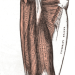 How Mechanism of Injury Affects Prognosis Following Hamstring Strain
 Acute Ankle Sprain: Brace Yourself…Or Should You?
 Manual Therapy for Inversion Ankle Sprains