INTRODUCTION
Acromioclavicular injuries are common in a variety of sports, particularly those which involve heavy contact or tackling. For example, Flik et al (2005) reported the incidence of AC injuries was the third most common in men’s ice hockey. Thus, this is a common injury. Whilst in many injury cases, such as acute presentations and the higher grade AC separations, the diagnosis can be quite obvious. However, many conditions of the shoulder present with very similar clinical presentations, and thus differential diagnosis can be challenging (Meyer et al 1990). Thus, this article examines the diagnostic accuracy for clinical examination tests for acromioclavicular joint pain.
This article is very similar in format to the articles:
PAXINOS TEST
- Sensitivity: 79%
- Specificity: 50%
- +ve LR: 1.58
- -ve LR: 0.42
Reference: Walton et al. (2004)
ACROMIOCLAVICULAR JOINT TENDERNESS
- Sensitivity: 96%
- Specificity: 10%
- +ve LR: 1.07
- -ve LR: 0.40
Reference: Walton et al. (2004)
O’BRIEN TEST
NB: Positive when the pain is reproduced over the AC joint area, not the anterior glenohumeral region (which would be more indicative of a SLAP tear).
- Sensitivity: 16 – 40%
- Specificity: 90 – 95%
- +ve LR: 1.60 – 8
- -ve LR: 0.63 – o.93
Reference: Walton et al (2004), Chronopoulos et al (2004)
HORIZONTAL ADDUCTION STRESS TEST
- Sensitivity: 77%
- Specificity: 79%
- +ve LR: 3.33
- -ve LR: 0.29
Reference: Chronopoulos et al (2004)
RADIOGRAPHS
i.e. Identified as showing any AC joint abnormalities e.g. joint space narrowing, marginal osteophytes, or subchondral cysts.
- Sensitivity: 41%
- Specificity: 90%
- +ve LR: 4.01
- -ve LR: 0.65
Reference: Walton et al (2004)
BONE SCAN
- Sensitivity: 82%
- Specificity: 70%
- +ve LR: 2.73
- -ve LR: 0.25
Reference: Walton et al (2004)
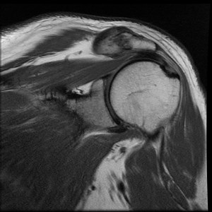
MAGNETIC RESONANCE IMAGING
- Sensitivity: 85%
- Specificity: 50%
- +ve LR: 1.70
- -ve LR: 0.3
Reference: Walton et al (2004)
CLINICAL IMPLICATIONS
This fairly obvious shows the poor diagnostic accuracy of physical examination tests and imaging for the diagnosis of AC joint pain. Some tests present with high sensitivity, however, this frequently corresponds with low specificity. Alternatively, the opposite is true. Being aware of tests with higher sensitivity will give negative results greater weight and high specificity will give positive results greater weight. For example: negative AC joint tenderness gives good negating evidence against AC joint pathology.
Thus, in this way you will require a battery of testing to ensure an accurate diagnosis. It is my intention that you could use this post as a reference for accurate clinical diagnosis of an AC joint injury. For increased accuracy you may require a Fagan’s Nomogram. As it always seems, be wary of these results; the articles cited below have a number of methodological faults.
What are your thoughts on accurate diagnosis of AC joints? Let me know in the comments or catch me on Facebook or Twitter
If you require any sports physiotherapy products be sure check out PhysioSupplies (AUS) or MedEx Supply (Worldwide)
REFERENCES
Chronopoulos E, Kim TK, Park HB, Ashenbrenner, D, McFarland EG. Diagnostic value of physical tests for isolated chronic acromioclavicular lesions. Am J Sports Med 2004 32: 655
Flik K, Lyman S, Marx RG. American collegiate men’s ice hockey: an analysis of injuries. Am J Sports Med. 2005;33:183-187.
Meyer SJ, Dalinka MK. Magnetic resonance imaging of the shoulder. Orthop Clin North Am. 1990;21:497-513.
Walton J, Mahajan S, Paxinos A, et al. Diagnostic values of tests for acromioclavicular joint pain. J Bone Joint Surg Am. 2004;86:807-812.
Related Posts


